
Neuroscience
Anatomically Accurate Brain Phantom
Experimental verification for neuromodulation
Virginia Commonwealth University researchers have developed an anatomically realistic 3-D brain phantom for the evaluation of neuromodulation techniques. Currently, there are no anatomically accurate brain phantoms that can experimentally verify neuromodulation methods such as transcranial magnetic stimulation (TMS), transcranial direct current stimulation (tDCS) and deep brain stimulation (DBS). This has caused experimental limitations on the ability to test different parameters of neuromodulation prior to clinical trials. The brain phantom developed by our researchers is anatomically realistic and created with conductive materials that allow for the testing of TMS, tDCS, and DBS along with various neuroimaging modalities.
The technology
This brain phantom is constructed by 3D printed shells and comprised and filled with a conductive material which is capable of mimicking the averaged conductivity of the grey matter (GM) and white matter (WM) in the brain. The unique design of these models allows for the testing of both neuroimaging and neuromodulation techniques, such as TMS, tDCS, and DBS in a manner similar to a clinical setting. This allows for a greater level of treatment planning and control prior to clinical trials. Additionally, these phantoms can be constructed for individual patients using their MRI data, so that testing can be tailored on an individual basis.
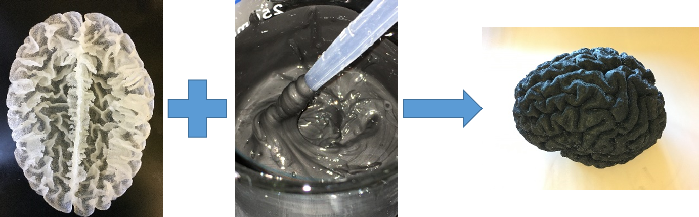
Figure 1. Constructing the brain phantom by the shell method where the conductive polymer is casted into the brain shell.