
Medical Imaging
Detecting Segmentation and Fracture in CT Images
A novel method to diagnose traumatic pelvic injuries
Traumatic pelvic injury is a severe yet common injury in the United States, often caused by motor vehicle accidents or fall. The severity and prognosis of these injuries are traditionally diagnosed through information contained in computed tomography (CT) images. Each pelvic CT scan includes a large number of slices, with each slice containing a large quantity of data that cannot be readily interpreted via a simple visual inspection. Therefore, a computer-assisted pelvic trauma decision-making system would be a valuable asset to help physicians improve their diagnostic decisions and quickly determine treatment plans. However, the design of such a system is a challenge as several factors need to be accounted for: limited resolution of medical images, variations in bone tissues, complexity of pelvic structures, and significantly different geometrical characteristics of fractures.
The technology
VCU researchers have developed a new hierarchical segmentation approach for the analysis of pelvic, and other bone, CT images. This approach is capable of automatically extracting multiple-level bone structures using a combination of anatomical knowledge and computational techniques: adaptive window creation, 2-D SWT application, masking, and boundary tracing. The proposed techniques then may use a template-based best-shape matching method that provides an entirely automated segmentation process (Figure 1). This would allow physicians to automatically and accurately detect fractures from segmented bones, and better determine the severity of pelvic injuries in patients.
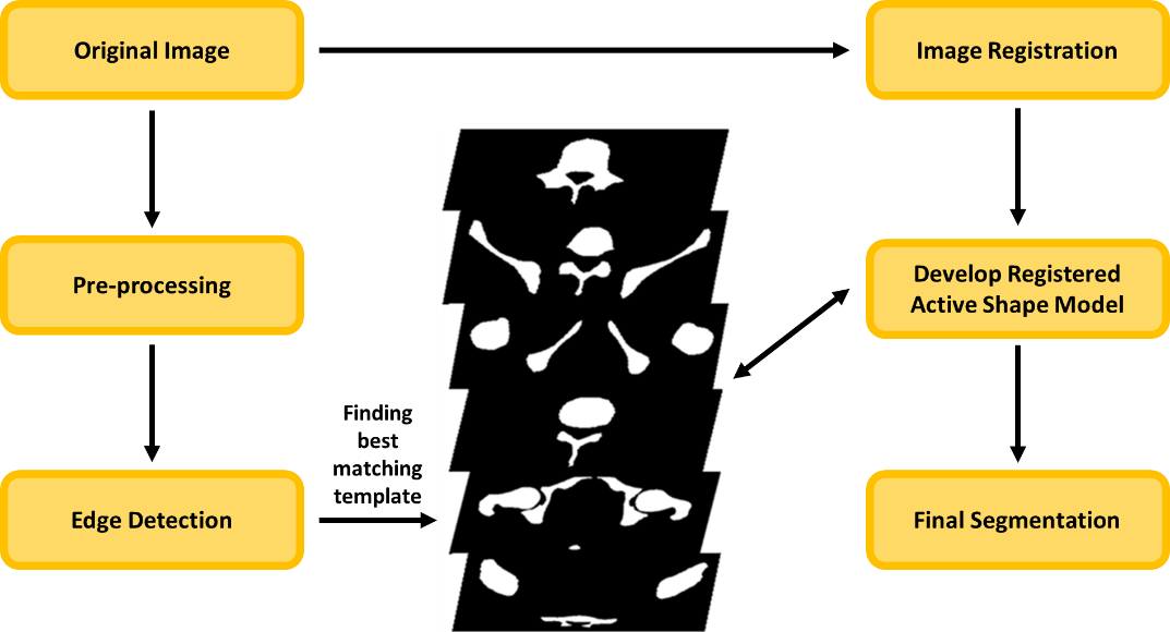
Figure 1. Schematic diagram of pelvic bone segmentation process