
Pelvis/femur modeling
Hip Analysis Suite
Automated diagnostic and preoperative software
Hip angle and size measurements are critical to hip replacement surgery success, and improper placement leads to abnormal biomechanical function. Current methods for pre-operative imaging rely solely on 2D imaging to develop models of these measurements. Doctors then use these models to diagnose alignments and provide aid during the operation of hip replacement surgery. However, these methods can lead to human error by the technician or surgeon and reduce likelihood of healthy patient outcome.
The technology
VCU researchers have developed an automated software package that provides 3D models and morphometric analysis of the femur and pelvis based off of a patient’s CT scan. Utilizing a novel algorithm, hundreds of landmarks are used to accurately estimate measurements and model the femur and pelvis of each individual patient. This information can then be applied to inform any hip reconstructive surgery or diagnose hip conditions. Using this software package, doctors or surgeons can build more accurate pre-operative hip models to increase hip replacement surgery success rates, and thus reducing healthcare costs associated with these surgeries.
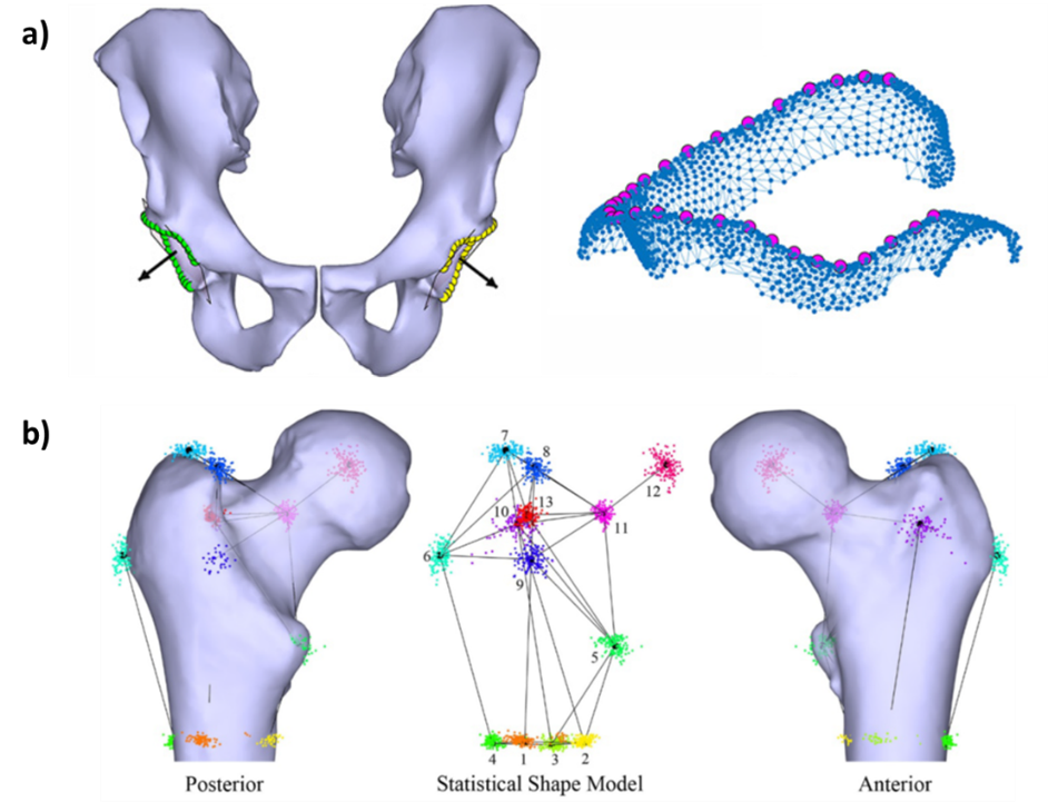
Figure 1a. Depiction of the automated detection of both acetabular orientation with normal vector (black arrow) designated and acetabular rim selection.
Figure 1b. Point distribution model showing locations of each point in the statistical shape model of the femora. A representative surface model is shown superimposed over the mean shape model in the anterior and posterior views.