
Software and informatics
Accurate Pelvic Injury Detection for X-Ray and CT Images
The technology
Pelvic fractures can be debilitating and even fatal if not treated correctly and in a timely manner. Current fracture imaging techniques are known to produce low resolution images and require some measure of manual interaction. Hence, there is a need for fully automated fracture imaging methods that are better able to analyze pelvic fracture images.
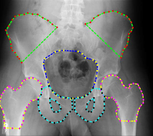
Researchers at VCU have developed methods to better assess the severity and location of pelvic fractures based on improved segmentation of X-ray or CT images. The methods have effective automatic initialization and produce a more accurate segmentation of images compared to existing methods, as shown in the figure. It also enables detection of hemorrhage in the pelvic region and separation of this from the detected bones.
Evidence has shown that features calculated from the known location of segmented bones - such as pubis bone gap - are strong indicators of pelvic injury severity. An experiment using these features for prediction of injury severity in trauma patients resulted in a prediction accuracy rate of 87.5%. This percentage is significant because other methods may not distinguish these diagnostic features with much accuracy or even at all.